In an evolving health landscape, emerging research continues to highlight concerns that could impact everyday wellbeing. Here’s the key update you should know about:
By seamlessly tracking brain growth from the womb into early life, researchers uncover when key brain regions surge and how subtle sex differences emerge before birth.
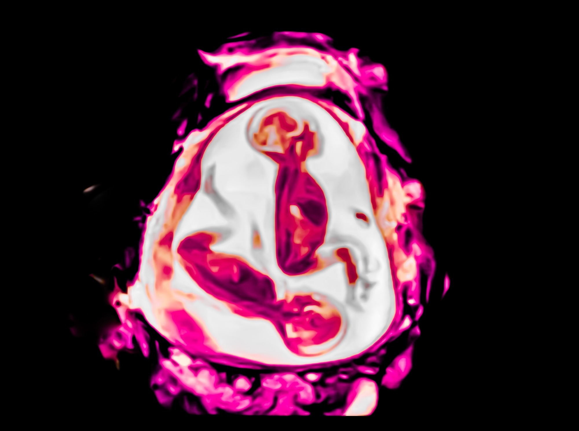
Study: Mapping brain growth and sex differences across prenatal to postnatal development. Image credit: Mapping brain growth and sex differences across prenatal to postnatal development/Shutterstock.com
The perinatal period is a key window of fetal neurodevelopment, encompassing both the prenatal and early postnatal periods. A recent neuroimaging study in the journal Scientific Reports explores brain growth during this period, using continuous prenatal-to-postnatal MRI data to better characterise how different brain tissues grow during this transition.
Hormones, environment, and early brain patterning
Brain growth in the perinatal period involves a host of processes. Various types of brain cells need to proliferate, migrate, and differentiate. Neuronal synapses must be established and circuits formed.
Most previous studies have used data from either the prenatal or the postnatal period alone. They provide only a discontinuous understanding of perinatal brain development. In particular, they do not follow the changes accompanying the tremendous transition from intrauterine to postnatal life.
Scientists have suggested that structural and functional neurodevelopment begins with basic sensory functions, followed by associative regions that mediate higher-order cognitive growth. Regional brain development is affected by maternal hormonal changes, shifts from placental to oral nutrition, and exposure to sensory stimuli, including social interactions.
Pregnancy complications and maternal drug use also have sex-specific biological effects on the developing brain. The male sex hormone testosterone, levels of which are approximately 2–2.5 times higher in male fetuses than in females, peaks at 14–18 weeks. This results in differential gene expression within placental and brain tissues, with downstream effects reported from around 18 weeks onwards.
Such deep-seated factors are assumed to contribute to later-life sex-dependent differences in the prevalence of various psychiatric and neurodevelopmental conditions, although such links remain indirect at this developmental stage.
The current study uses publicly available large datasets to address past data scarcity and maps separate regional and overall growth trajectories in the brain during the perinatal period, using both absolute and proportional brain volumes.
Developing Human Connectome Project
The study used perinatal magnetic resonance imaging scans from the Developing Human Connectome Project. The researchers had access to 798 scans from 699 people, 263 of which were prenatal and the rest neonatal. Ninety-seven participants had longitudinal data, with 78 undergoing both prenatal and postnatal scans, mostly postnatally. There were 380 males. Both absolute and proportional brain volumes were estimated.
The study aimed to model brain development, total and proportional, during the perinatal period. It also sought to compare brain volumes between 21 and 45 weeks from conception, in males and females.
Study findings
Total brain volume
The study shows that total brain volume increased during the perinatal period.
This agrees with earlier prenatal neuroimaging studies indicating the highest brain growth velocity beginning in the late second trimester, with growth rates slowing during the early postnatal weeks when postnatal age at scan is accounted for.
White matter volume
At mid-gestation, most brain growth occurred in the white matter, which accounted for a larger share of brain volume before 35 weeks. Its proportion to total brain volume progressively dropped over the perinatal period.
This is concordant with prior research showing peak white matter growth contribution at 29–30 weeks. All major white matter tracts are formed at term, and some, like the thalamo-cortical tract, at the start of the third trimester. Moreover, white matter injury is among the defining differences between babies born preterm and at term, as reported in previous studies, rather than measured directly here.
Gray matter volume
Gray matter grew most rapidly in late pregnancy and postnatally. It comprised a greater volume of the brain after 35 weeks. This may help support the increasing integration of sensorimotor and cognitive functions during the perinatal period.
These findings are consistent with earlier studies showing a relatively modest increase in white matter volume over the first year of postnatal life, compared with much larger increases in gray matter, although these postnatal percentages were not measured in the current study. White matter volume peaks only in adulthood, whereas gray matter peaks in childhood.
Subcortical gray matter
Subcortical gray matter includes the amygdala, hippocampus, basal ganglia, and thalamus. The study revealed that its growth began earlier than cortical gray matter, peaking in the third trimester. Coupled with postnatal deceleration, this likely reflects an initial period of rapid expansion that slows as regional size increases.
Very rapid, accelerating growth of the cerebellum was observed in contrast to more linear hippocampal growth throughout the perinatal period.
Previous research has shown lower subcortical volumes, especially in the basal ganglia and thalamus, in very preterm infants compared to term infants of comparable postnatal age. These differences have been correlated with adverse cognitive, behavioral, and motor outcomes, but were not directly assessed in this study.
Among all brain regions, the cerebellum shows the fastest growth in later pregnancy and early neonatal life, which may relate to the early development of motor coordination. Perturbations of this process have been associated with poor cognitive, motor, social, and emotional outcomes in prior work.
The hippocampus has been shown to mature the slowest among brain regions in postnatal life, a pattern thought to reflect its involvement in increasingly complex higher-order brain functions.
Sex differences in brain growth
In this study, brain volume was higher in males at all ages, and males showed greater age-related increases in absolute brain volume, although overall growth trajectories were broadly similar between the sexes.
Despite this, gray matter growth rates in specific regions were accelerated in males. These included parts of the right inferior temporal gyri and left parietal lobes.
Notably, the growth of the left anterior cingulate gyrus, linked to social cognition, slowed towards the end of the perinatal period in males. Its consistently larger size in females throughout development may be relevant to previously reported early and sustained advantages in this domain. Alternatively, it could reflect the larger proportional size of this region in females, rather than faster absolute growth.
Importantly, when regional volumes were analysed as proportions of total brain volume, fewer sex-by-age interactions were observed, suggesting that some apparent regional differences may be driven by overall differences in brain size between sexes.
Sensitivity analyses of neonatal scans with respect to postnatal age confirmed a general slowing of growth, except for subcortical structures.
Methodological differences complicate fetal–neonatal comparisons
This large study used scans beginning immediately after birth, capturing early neonatal brain growth. However, its limitations include:
- Paucity of neuroimaging data
- No scans before 21 weeks
- Few second-trimester scans
- Few scans after 37 weeks hinder the understanding of growth events in the final part of pregnancy
- Longitudinal data in only 14 % of the sample
- Different methods in fetal vs neonatal scans, potentially affecting volume measurements
- No fetal body size measurements, preventing comparisons between sex-specific overall growth and brain growth
- Only one of several brain growth measurements was captured
In addition, cerebrospinal fluid and intracranial volume trajectories were modelled in the study but were not central to its primary conclusions and are not discussed in detail here. Future work with longer follow-up is necessary to validate these findings.
A continuous view of perinatal brain growth
This study is the first to present perinatal trends in brain volume growth, stratified by sex and spanning the early postnatal weeks. Put together, these findings suggest a rapid establishment of key neuronal circuits and networks during fetal life, followed by slower maturation and myelination of these connections after birth.
It also emphasizes a shift in growth trajectories toward term, perhaps in preparation for the impending birth. The observed sex-specific differences in brain growth are consistent with, rather than definitive evidence of, the timeline of prenatal shifts in sex hormone levels.
These findings underline the need to map early brain growth using prenatal and postnatal imaging. In particular, they shed light on sex-specific brain growth patterns that could potentially be combined with future longitudinal and clinical studies to help explore possible mechanistic links to neurodevelopmental and psychiatric conditions.
Journal reference:
-
Khan, Y. T., Tsompanidis, A., Radecki, M. A., et al. (2026). Mapping brain growth and sex differences across prenatal to postnatal development. Scientific Reports. DOI: https://doi.org/10.1038/s41598-025-33981-w. https://www.nature.com/articles/s41598-025-33981-w