In an evolving health landscape, emerging research continues to highlight concerns that could impact everyday wellbeing. Here’s the key update you should know about:
New research reveals how common respiratory viruses can flip dormant breast cancer cells back into growth mode, uncovering an immune-driven pathway that heightens relapse risk and pointing to new prevention strategies.
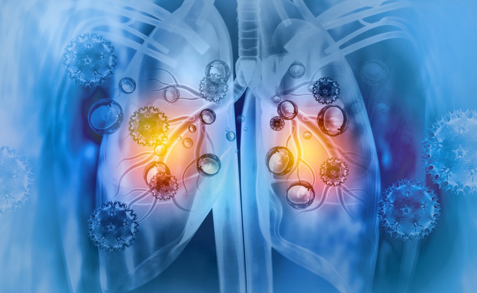
Study: Respiratory viral infections awaken metastatic breast cancer cells in lungs. Image Credit: crystal light / Shutterstock
In a recent study published in Nature, an international team of researchers showed that respiratory viral infections awaken dormant breast cancer cells in the lungs.
Breast cancer is the most prevalent cancer in females and the second leading cause of cancer-related deaths in the United States (US). Disseminated cancer cells (DCCs) can remain dormant for years after initial remission before metastatic relapse. The tumor microenvironment and cell-intrinsic factors determine whether metastatic cells progress or remain dormant. Notably, microenvironmental disturbances can be sufficient to increase metastasis.
Respiratory viral infections are common, with seasonal flu affecting over one billion people yearly. These infections are usually associated with pulmonary inflammation along with an increase in inflammatory cytokines (interferons [IFNs] and interleukin 6 [IL-6]) and expansion of immune cells, such as macrophages, T cells, and neutrophils. Such inflammatory mechanisms have been reported as regulators of metastatic processes.
The study and findings
In the present study, researchers examined the effects of respiratory viral infections on breast cancer dormancy in mice. First, they used a mouse model of breast DCC dormancy, MMTV-Her2, to explore the effects of influenza A virus (IAV) on the awakening of dormant DCCs. Mice were infected with a sub-lethal dose of IAV; both MMTV-Her2 and wild-type mice showed comparable inflammatory response and viral clearance kinetics.
Lungs were harvested at several time points and assessed for the abundance of human epidermal growth factor receptor 2-positive (HER2+) cells. Before infection, a few isolated DCCs or clusters of DCCs were detected. Nevertheless, metastatic burden increased by up to 1,000-fold between three and 15 days post-infection (dpi). The number of HER2+ cells remained elevated at 28 and 60 dpi and was still detectable nine months later.
There were no changes in Ki67+HER2+ cells in mammary glands, and qPCR of blood samples showed no increase in circulating cancer cells, suggesting that the increase in HER2+ cells in the lungs was not derived from elevated seeding of cancer cells in mammary glands.
Further, the team observed a significant increase in HER2+ cells expressing Ki67 at 3 dpi. Although Ki67-expressing HER2+ cells decreased by 15 dpi, the number of these cells remained elevated at 60 dpi compared to baseline.
Dormant DCCs maintain a mesenchymal-like state (vimentin-positive) and undergo an epithelial shift (epithelial cell adhesion molecule-positive [EpCAM+]) during dormancy exit. Most dormant DCCs in uninfected lungs were vimentin+. While the percentage of vimentin+ HER2+ cells was not affected early in infection (3 to 6 dpi), it decreased to 50% by 9 dpi and less than 20% by 28 dpi. In contrast, a fraction of HER2+ cells showed EpCAM expression by 3 dpi.
Moreover, while most HER2+ cells lost EpCAM positivity after 6 dpi, the percentage of EpCAM+ HER+ cells remained elevated. Thus, IAV infection induced a transient epithelial shift, creating a unique hybrid and proliferative phenotype that retained some mesenchymal marker expression, allowing dormant DCC awakening.
RNA-seq analyses showed activation of inflammatory (IL-6–JAK–STAT3), angiogenesis, and extracellular matrix–remodelling pathways, including collagen crosslinking and metalloproteinase activity, which are known to support tumour growth.
The authors also reported shifts in the tumour microenvironment, including extracellular matrix changes and angiogenic signalling, that could help sustain awakened DCCs. The team also noted the activation of the IL-6 signaling pathway in DCCs post-infection. Further investigations indicated that infection-triggered IL-6 was key in mediating initial dormant DCC awakening.
The researchers identified a two-phase process: first, IL-6 drives the switch from a mesenchymal to a hybrid phenotype and fuels rapid expansion; later, after T-cell recruitment, CD4+ T cells sustain the awakened DCC population. During this second phase, CD4+ cells partly maintain DCCs by suppressing CD8+ immune responses.
Gene expression profiling revealed that CD4+ cells in tumour-bearing mice had reduced mitochondrial content, a bias toward a memory phenotype, and lower effector function, further limiting CD8+ cytotoxicity.
The study also found that depleting CD4+ cells restored CD8+ cell mitochondrial content and effector activity, leading to more effective elimination of DCCs.
Next, the team investigated whether coronavirus disease 2019 (COVID-19) can awaken dormant DCCs. To this end, a mouse-adapted severe acute respiratory syndrome coronavirus 2 (SARS-CoV-2) strain (MA10) was used. MA10 infection triggered the production of IFNα and IL-6 in the lungs.
Besides, MA10 infection resulted in a notable increase in HER2+ cells by 28 dpi. Moreover, there was a stepwise increase in the number of HER2+ cells and Ki67+HER2+ cells following MA10 infection, with reductions in vimentin positivity and transient increases in EpCAM positivity. Consistently, these changes required IL-6, as changes associated with MA10 infection were significantly reduced in IL-6 knockout mice.
Further, the researchers analyzed data from the United Kingdom Biobank (UKB) to assess whether a positive SARS-CoV-2 test was associated with a higher risk of mortality among cancer survivors. In a UKB population followed up until December 2022, which included 4,837 individuals with a cancer diagnosis before 2015, 413 deaths were recorded. These included 115 and 298 deaths, those who tested positive and negative for SARS-CoV-2, respectively, yielding an odds ratio (OR) of 4.5.
Even after excluding COVID-19-attributed deaths, test-positive individuals still had higher mortality, with an OR of 2.56. There was a nearly two-fold increase in cancer mortality (OR: 1.85) in test-positive individuals compared to test-negative participants.
The data showed that the association was strongest in the months immediately after infection and weakened over time, mirroring the early rapid expansion of DCCs seen in the mouse models. The team observed elevated risks for all-cause, non-COVID-19, and cancer mortality among participants who tested positive for SARS-CoV-2 compared to those who tested negative.
Finally, the Flatiron Health Database was used to evaluate whether females with breast cancer experienced a higher risk of metastatic progression to the lungs after COVID-19. Females with COVID-19 after breast cancer diagnosis had a hazard ratio of 1.44 for subsequent diagnosis of metastatic breast cancer, adjusted for age, race, and ethnicity. After additional adjustment for breast cancer subtype and comorbidities, the hazard ratio was 1.41 and no longer statistically significant, although the direction of effect was consistent.
Conclusions
The results indicate that respiratory viral infections promote awakening and expansion of dormant cancer cells. An IL-6-dependent switch from a mesenchymal state to a hybrid phenotype promotes expansion, followed by the establishment of CD4+ niches that inhibit DCC elimination.
These niches also impair CD8+ antitumour activity by altering immune cell metabolism and effector potential. Other immune cell populations, including macrophages, also showed phenotype shifts toward a tumour-supportive state.
Overall, these data reveal how pulmonary viral infections elevate cancer recurrence risk, with both mouse and human data showing the greatest risk in the early period after infection, underscoring the need for strategies to alleviate the increased risk of associated metastatic progression.

