Scientists Map Aging Brain in Unprecedented Detail, Revealing Clues to Alzheimer’s and More
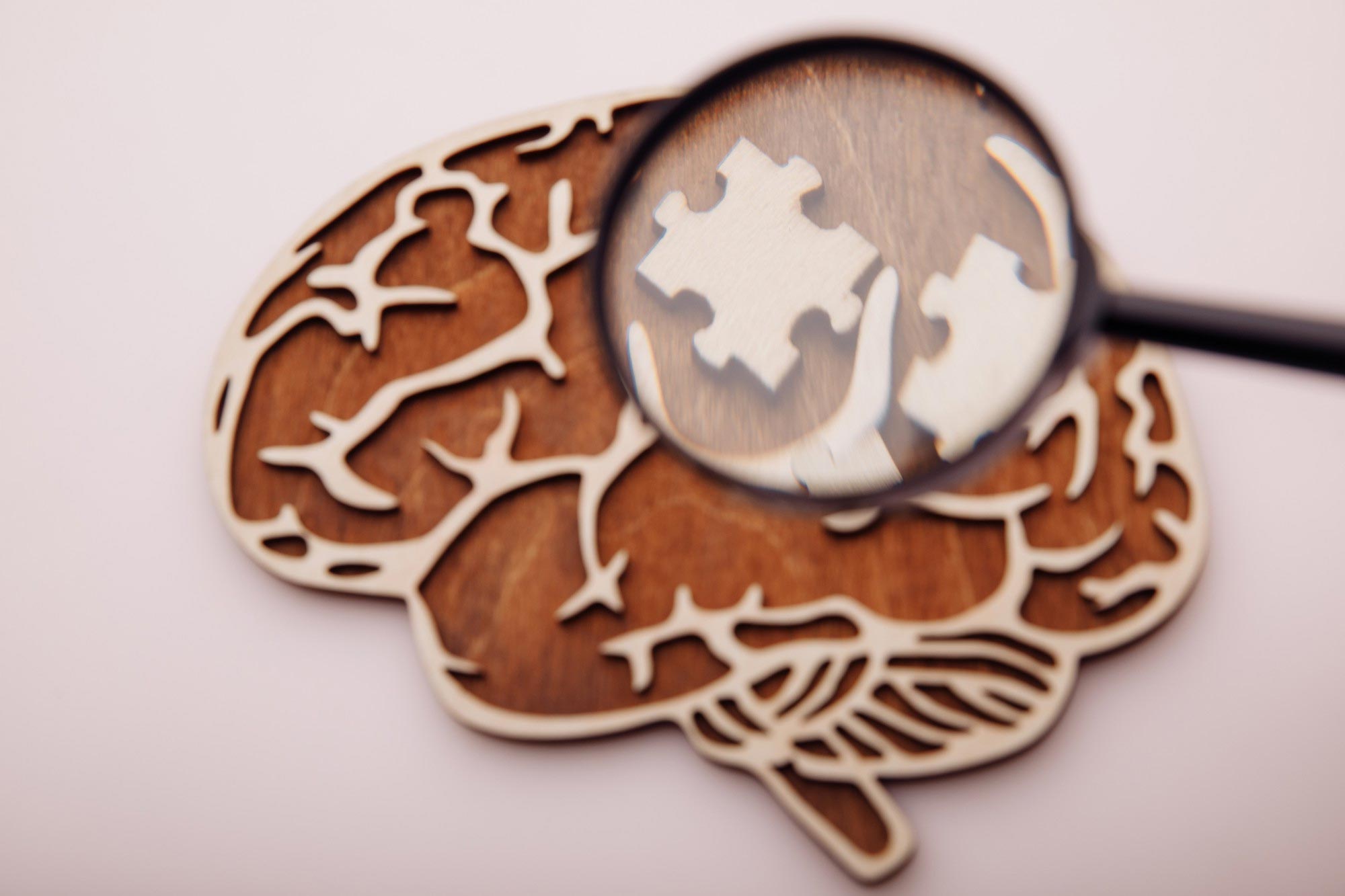
A new single-cell atlas shows how epigenetic changes reshape brain cells during aging, revealing genomic instability, regional differences, and potential biomarkers of brain aging.
More than 57 million people worldwide are currently living with neurodegenerative diseases. These conditions include Alzheimer’s disease, Parkinson’s disease, ALS, and others. Researchers expect the number of cases to double roughly every 20 years. Aging is known to be one of the strongest risk factors for these disorders, yet scientists are still working to understand exactly how aging drives changes in the brain.
One of the most important biological processes involved is epigenetic change. This refers to chemical markers that sit on top of DNA and influence how genes are turned on or off over time. Scientists at the Salk Institute have now produced the most detailed single-cell atlas so far of epigenetic changes in the aging mouse brain.
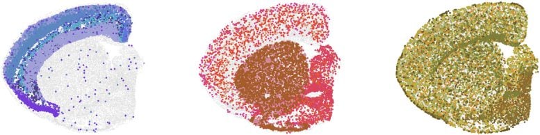
Their work shows how DNA methylation, genome organization, and gene activity evolve across different brain regions and cell types as animals grow older. The dataset spans eight brain regions and includes 36 different brain cell types. More than 200,000 single cells were analyzed for methylation and chromatin conformation, and nearly 900,000 cells were mapped using spatial transcriptomics.
Scientists Release Public Atlas of Aging Brain Epigenetics
The dataset has already revealed distinct epigenetic differences between younger and older brains. It also enabled researchers to build new deep learning models that can forecast how gene activity shifts with age. The findings were published in Cell on March 11, 2026. The atlas is now publicly accessible through Amazon Web Services (AWS) and Gene Expression Omnibus (GEO). Scientists expect it to serve as a foundational reference for interpreting human brain datasets, including those produced by the National Institute of Health’s Brain Research Through Advancing Innovative Neurotechnologies (BRAIN) Initiative.
“Age-related brain changes, particularly in regions critical for attention, memory, emotion, and motor functions, severely impact life quality,” says co-corresponding author Joseph Ecker, PhD, professor and holder of the Salk International Council Chair in Genetics at Salk and a Howard Hughes Medical Institute Investigator. “By mapping how the epigenome changes across individual brain cell types as animals age, we now have a framework for understanding how aging reshapes the brain at the molecular level. This resource should help researchers pinpoint mechanisms that contribute to neurodegenerative disease.”
Epigenetic Hallmarks of Aging and Methylation Changes
Aging is linked to four major molecular features: chronic inflammation, mitochondrial dysfunction, genome instability, and epigenetic alterations. Growing evidence suggests that the epigenome plays a central role in driving the aging process. One specific epigenetic modification, known as methylation, has been closely connected to brain function, behavior, and neurological disease. If researchers can better understand how methylation patterns shift with age, they may eventually develop strategies to reverse harmful changes and restore cellular health.
However, collecting meaningful methylation data is challenging. Studying only some brain cells does not capture the full complexity of the organ. The brain contains many specialized regions and diverse cell populations, all of which must be examined to gain a complete understanding of aging.
“The brain is so interconnected, with different regions controlling different functions and aging at different speeds at the cell type level,” says co-corresponding author Margarita Behrens, PhD, a research professor at Salk. “We can see how interconnected the brain is in conditions like Parkinson’s, where the death of one group of neurons spirals into an entire circuit malfunctioning and then the tremors and cognitive effects we see in patients. So, the importance of having a cell type-specific understanding of aging will bring more granular knowledge that will expand therapeutic possibilities.”
Single-Cell Multi-Omic Mapping of the Aging Brain
Traditional bulk analyzes combine signals from many cells, which hides differences between cell types. Single-cell approaches overcome this limitation, allowing researchers to examine individual cells in detail. With this in mind, the Salk team set out to build one of the most extensive single-cell multi-omic brain datasets available.
In addition to measuring DNA methylation, the researchers examined chromatin conformation, which describes the three-dimensional arrangement of DNA inside the cell nucleus. They also applied advanced spatial transcriptomics methods to track gene expression while preserving the original position of cells within brain tissue.
“What makes this work innovative is, above all, its spatial dimension,” explains first author Qiurui Zeng, a graduate student in Ecker’s lab. “Spatial resolution reveals which regions and local microenvironments are most vulnerable to aging, how cell-type composition shifts across brain areas over time, and how neighboring cells may influence one another’s aging trajectories. The scale of the spatial dataset—nearly 900,000 spatial transcriptome cells—is itself unprecedented for a longitudinal aging study.”
Using a mouse model of aging, the researchers gathered methylation data from 132,551 individual brain cells and combined methylation and chromatin conformation information from another 72,666 cells. Altogether, the dataset includes 36 major cell types. The complete dataset was released on AWS and GEO in December 2025.
Cloud-Based Open Data Accelerates Brain Aging Research
Placing the dataset on AWS makes it widely accessible while removing the heavy computational barriers typically required to handle large biological datasets. Hosting the nearly 900,000 spatially mapped cells in the cloud allows scientists around the world to explore the data without needing specialized infrastructure.
“The AWS Open Data program covers storage costs and places this dataset alongside other major neuroscience resources like the Allen Brain Atlas and the Seattle Alzheimer’s Disease Brain Cell Atlas, making it part of an interconnected ecosystem of publicly accessible brain data,” adds Zeng. “Researchers in aging, neurodegeneration, and spatial genomics can build on this resource immediately, accelerating the pace of discovery well beyond what a single lab could achieve.”

The methylation results revealed that age-related changes were more pronounced in non-neuronal cells. The researchers also observed that transposable elements, often referred to as ‘jumping genes,’ lose DNA methylation as cells age. This suggests that sections of the genome that are usually inactive may become more active in older brains.
Jumping genes are repetitive DNA sequences that make up roughly half of the human genome. When these sequences become active, they can disrupt normal cellular function and contribute to age-related decline. The findings support the idea that epigenetic shifts play a role in cellular dysfunction associated with aging.
Chromatin Structure Changes Reveal New Brain Aging Biomarker
The chromatin conformation data uncovered additional structural changes linked to aging. In particular, the team identified a potential biomarker of brain aging. They observed stronger signals at topologically associating domain (TAD) boundaries and increased accessibility at nearby CTCF binding sites.
Genomes organize their vast amount of information using structures called TADs. These are segments of DNA that interact closely with each other. The protein CTCF binds to the edges of these domains and helps maintain their structure.
Researchers then integrated the spatial transcriptomics results. Nearly 900,000 cells were analyzed to compare how aging unfolds across various brain regions and cell types.
“The same cell type ages differently depending on its location; for instance, non-neuronal cells in the back of the brain show more inflammation than those in the front parts,” says Zeng. “This data really underscores the variability in aging even among the same cell type, emphasizing the importance of cell and brain region-level specificity in unraveling the complexities of aging.”
Deep Learning and the Future of Brain Aging Models
The dataset has already produced valuable insights. The researchers created deep learning models capable of predicting gene expression patterns using future multi-omic epigenetic data. This approach could eventually support the development of a virtual model that simulates how the brain ages.
The atlas is now openly available for scientists around the world. By sharing the resource widely, the researchers hope global collaboration will accelerate discoveries related to brain aging and neurodegenerative disease.
Reference: “Cell-type-specific transposon demethylation and TAD remodeling in aging mouse brain” by Qiurui Zeng, Wenliang Wang, Wei Tian, Amit Klein, Anna Bartlett, Hanqing Liu, Joseph R. Nery, Rosa G. Castanon, Julia Osteen, Nicholas D. Johnson, Wubin Ding, Huaming Chen, Jordan Altshul, Mia Kenworthy, Cynthia Valadon, William Owens, Zhanghao Wu, Maria Luisa Amaral, Nathan R. Zemke, Yuru Song, Cindy Tatiana Báez-Becerra, Silvia Cho, Chumo Chen, Jackson Willier, Stella Cao, Jonathan Rink, Jasper Lee, Ariana Barcoma, Jessica Arzavala, Nora Emerson, Yuancheng Ryan Lu, Bing Ren, M. Margarita Behrens and Joseph R. Ecker, 11 March 2026, Cell.
DOI: 10.1016/j.cell.2026.02.015
The work was supported by the National Institutes of Health (5R01AG066018-05, RRID: SCR_014839, CCSG P30 CA014195, S10-OD023689, S10 OD034268) and Howard Hughes Medical Institute.
Never miss a breakthrough: Join the SciTechDaily newsletter.
Follow us on Google and Google News.
Source link