In an evolving health landscape, emerging research continues to highlight concerns that could impact everyday wellbeing. Here’s the key update you should know about:
In this interview, News Medical-Life Sciences speaks with Teemu Miettinen, cell biologist at the Massachusetts Institute of Technology, about research into measuring intracellular water and its impact on cellular function and disease. Drawing on new methodology, Miettinen explains how water content influences biochemical reactions, cell growth, and emerging research in cancer, immunology, and beyond.
Can you please introduce yourself and your role at MIT?
My name is Teemu Miettinen. I am originally from Finland, where I completed my undergraduate and Master’s studies, before moving to Scotland for my PhD, which was focused on cell size regulation.
For the past decade, I have been working at the Massachusetts Institute of Technology, where I conduct research to understand how cells regulate their size, growth, and composition.
By training, I’m a cell biologist and a biochemist, but my work is centered on fundamental cell biology and on developing new methods to measure biophysical properties of cells.
One of the key questions driving my research is how cell size actually matters. Cells in the human body can vary enormously in size, spanning up to seven orders of magnitude, and there is size variability even within any given cell type. Yet, we still do not fully understand how this variation impacts cellular function, like a cell’s capacity to grow. What we do understand much better is the molecular side, particularly that the concentration of molecules inside cells is critical for biological processes. And these concentrations sometimes change with cell size.
This is where cell composition becomes important. My work aims to understand what a cell is fundamentally made of, and a central part of that question is a very basic yet surprisingly understudied question: how much water is actually inside a cell and how this is regulated.
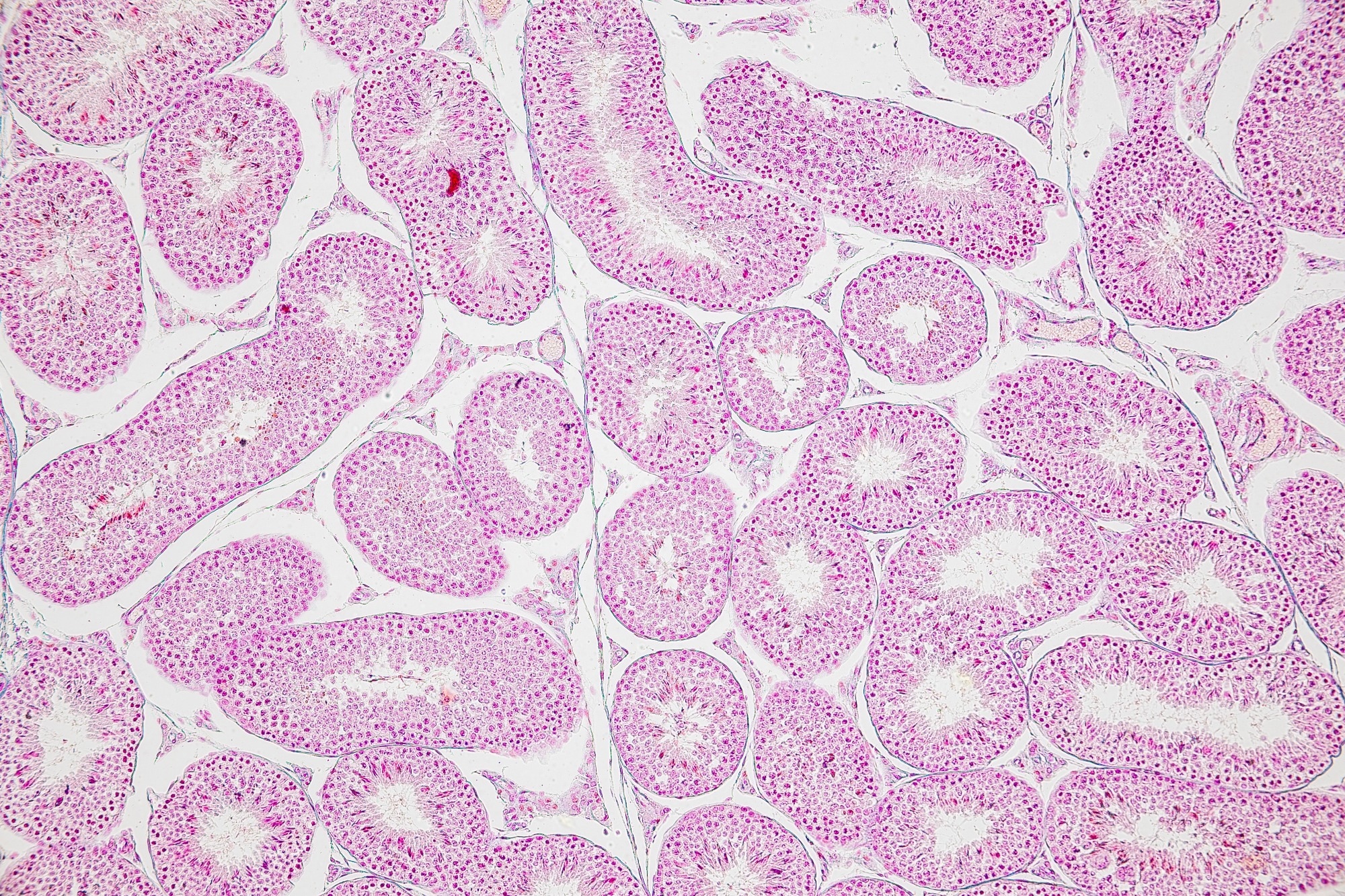
Microscopic view of tissue composed of densely packed cells, highlighting the complex organization and structure that underpin cellular function and biological processes. Yet, most of such tissue mass is water, and the amount of water is critical for the tissue function. Image credit: Sinhyu Photographer/Shutterstock.com
Why is intracellular water content so important for cell function?
Water content is fundamental because it directly determines the concentration of every molecule inside a cell. Since biochemical reactions depend on the concentrations of enzymes and substrates, altering water concentration alters reaction rates throughout the cell.
As water content decreases, concentrations increase, which can accelerate reactions up to a point. However, there is also an opposing effect. As the cell becomes more crowded with molecules, diffusion slows down, particularly for larger molecules and macromolecular complexes.
This creates a balance. Too little water leads to crowding and slower diffusion, while too much water dilutes molecules and reduces reaction efficiency. As a result, each cellular process may have its own optimal water content, depending on its sensitivity to concentration and diffusion.1
What challenges have historically limited the measurement of cellular water content?
Traditional methods for measuring water content are not well-suited to biological systems. For example, weighing and drying samples requires large quantities of material and destroys the samples. Other approaches, such as Raman spectroscopy-based imaging, can detect water but struggle to distinguish between water inside and outside the cell, especially since cells are typically surrounded by aqueous environments.
Additionally, imaging methods can be limited when working with thicker or more complex biological samples, making it difficult to obtain precise measurements in physiologically relevant systems. This is why we needed an approach that does not rely on indirect or destructive techniques, but instead measures fundamental physical properties, such as mass, more directly.
What new method have you and your team developed to overcome these limitations?
We developed a method based on measuring the buoyant mass of cells in different solutions. The approach involves weighing a sample multiple times in different fluids, including one containing heavy water, also known as deuterium oxide.
To do this, we use an inertial-sensing system.2 In simple terms, we flow the sample through a small vibrating metal tube. As the tube vibrates, it has a natural resonance frequency, and when a cell or organoid passes through it, even a tiny increase in mass causes a measurable shift in that frequency. It’s very similar to how a guitar string produces a lower pitch when it becomes heavier. By precisely tracking these frequency changes, we can determine the sample’s buoyant mass with high sensitivity.
By repeating the measurement in different solutions, we can back-calculate the water content of the sample in absolute and relative (volume/volume) terms, as well as other physical properties, such as dry mass.
Importantly, this method does not destroy the sample and allows us to study more complex and physiologically relevant biological systems, such as tumor spheroids and organoids.
How does your technique differ from previous technologies in terms of accessibility?
Our earlier work studying intracellular water content has relied on highly specialized single-cell mass sensors that are extremely powerful, but require custom-built components, significant expertise to operate, and are not something most labs can realistically implement.
That’s actually something I’ve always found a bit frustrating. I’ve been lucky to work in a place where we can do these kinds of measurements, but at the same time, it means most other biologists simply can’t.
With this new approach, we wanted to move away from that. The core idea was to build something that is much more accessible in terms of hardware. In principle, all the components we use are commercially available. You can source them from standard suppliers, often even from manufacturers outside the highly specialized scientific sector.
Of course, it still requires engineering know-how to assemble and optimize the system properly, especially for controlling vibration and ensuring measurement precision. But there’s nothing fundamentally custom or prohibitively expensive about the hardware. The hope is that this approach makes it much more feasible for other labs to adopt water-content measurements and to start exploring these questions more broadly.
How does intracellular water content relate to cancer biology?
Every reaction in the cell likely has its own optimal water content. We already know from a range of studies that if you change the water content of a cell, for example, by adjusting the external osmolarity, you can directly affect how fast that cell grows. You can pull water out of the cell or allow more to flow in, and that alone will change its growth rate. A lot of this work hasn’t necessarily been done in cancer models specifically, but the same principles apply across biology, even in very simple systems like bacteria.
So one fairly straightforward hypothesis is that cancer cells have adopted a different intracellular water content to support their high growth rates.
We already see something similar in the immune system. For example, T cells that are not actively growing tend to have relatively low water content, around 60 %. But when they become activated and start proliferating, their water content increases substantially, going up to 80 %.
So there is a clear link across multiple systems between water content and growth. We would expect cancer cells to follow the same kind of relationship. If that is the case, then it also suggests something more practical. If you can start to perturb the water content of a cancer cell, you are likely going to perturb its growth as well.
Beyond cancer, what other areas could benefit from this research?
We are currently looking quite a lot at the immune system, because that’s another area where we know water content regulation plays a role in growth and function. But more broadly, you can almost pick any biological system, and water content is likely to be relevant.
What’s exciting about this method is that it works across very different size scales. We’ve shown it in tumor spheroids, but in principle, you could go much further. You could imagine measuring whole organisms, something like a tardigrade, which can survive extreme dehydration, or even small animals like C. elegans. We’re also developing miniaturized versions of the method, so it works for single cells. In fact, we have already found radical water content regulation in some marine algae, where water regulation might influence cell buoyancy and photosynthesis.3
And because the method is non-invasive, you can measure the same sample repeatedly over time. That means you can start tracking how water content changes during growth or in response to different conditions or perturbations, which hasn’t really been possible before.
How important was interdisciplinary collaboration in this work?
This research would not have been possible without an interdisciplinary approach. I’m working as a part of a lab that is, at its core, an engineering lab. It’s focused on cancer, but it approaches it by building new tools. And I come in as a cell biologist with questions that have been very difficult to answer using existing methods.
So you really need both sides. You need people who can build measurement systems, and you need people who understand the biology well enough to ask the right questions.
And it’s not just one type of engineering either. There’s mechanical engineering, electrical engineering, and then you also have to think carefully about data analysis, because when you’re developing a completely new method, there isn’t really an established pipeline to follow.
I think a good example of this interdisciplinary work is that most cell biologists wouldn’t think about weighing a cell in heavy water, and engineers working with biological systems wouldn’t necessarily arrive at that idea either. It really comes from combining those different perspectives. And, of course, there are many people that have contributed to this over the years.
How do you see this technique evolving in the future?
One of the most exciting aspects is that the method does not destroy the sample. That immediately opens up many possibilities.
You can start combining water content measurements with other types of analysis. For example, you could perform high-content imaging alongside it, or examine how the structure of a tumor spheroid relates to its water content. You can also imagine applying this in more automated settings, like in drug development, where you’re testing how different compounds influence cellular water content.
And beyond that, you could combine it with omics approaches, for example, looking at proteomic composition at the same time.
So really, the key point is that once the sample remains intact, you’re no longer limited to a single measurement. You can start layering different types of data on top of each other, which opens up many directions for how this could develop going forward.
We are exploring many future directions for this work, but I hope others can join us in this exploration now that we finally have more tools to study water content.
Where can readers find more information?
About Teemu Miettinen
Teemu P. Miettinen, PhD, is a Research Scientist at the Koch Institute for Integrative Cancer Research at the Massachusetts Institute of Technology. He leads research seeking to understand how cell size, growth, and composition are regulated and interconnected. The research utilizes various model systems ranging from human cancer and immune cells to single-celled plankton. He also works jointly with the laboratory of Prof. Scott Manalis, developing novel biophysical measurements for biomedical applications.
His current research is funded by NIH, he co-holds two patents on measurement technologies, and he has authored several dozen research papers on the regulation of cell size, growth, and composition.
Source:
For other research conducted by Teemu Miettinen, see here.
Journal references:
- Huang J, Ferrell J (2026). How does cytoplasmic crowding affect reaction rates? Molecular Cell 86: 9–23. DOI: 10.1016/j.molcel.2025.12.007. https://www.cell.com/molecular-cell/abstract/S1097-2765(25)00981-5
- Katsikis G, et al. (2026). Inertial sensing of water content in tumor spheroids. Science Advances 12: eaeb1451. DOI: https://doi.org/10.1126/sciadv.aeb1451. https://www.science.org/doi/10.1126/sciadv.aeb1451
- Wu Y, Kouadio VK, Usherwood TR, Li J, Bisher M, Aurora R, et al. (2025). Diverse biophysical and molecular mechanisms drive phytoplankton sinking in response to starvation. PLOS Biology 23(11): e3003508. DOI: https://doi.org/10.1371/journal.pbio.3003508. https://journals.plos.org/plosbiology/article?id=10.1371/journal.pbio.3003508