For more than ten years, Evan Economo’s lab has relied on micro CT scanners to image insect specimens. These X ray scans allow scientists to examine the physical structure and form of insects, an area of research known as morphology. Although the technique provides extremely detailed 3D data, it is expensive and slow.
“One limitation is that you can get this rich 3D dataset, but it could take 10 hours to scan one specimen,” explained Economo, chair of the University of Maryland’s Department of Entomology and holder of the James B. Gahan and Margaret H. Gahan Professorship.
In a study published in the journal Nature Methods on March 5, 2026, Economo and colleagues tested a new approach designed to dramatically speed up the process. The project brought together researchers led by Economo and Thomas van de Kamp at the Karlsruhe Institute of Technology (KIT) in Germany. Their team combined a Synchrotron particle accelerator, X ray imaging, robotics, and artificial intelligence (AI) to generate interactive digital reconstructions representing 800 ant species.
Together, these technologies allowed the scientists to scan specimens far more quickly and convert raw imaging data into detailed 3D models.
“We’ve estimated that if we were to carry out this project with a lab-based CT scanner, it would take six years of continuous operation,” said Julian Katzke, the study’s first author and a graduate of Economo’s lab at the Okinawa Institute of Science and Technology (OIST) in Japan. “With the setup at KIT, we scanned 2,000 specimens in a single week.”
The effort, known as Antscan, could guide future large scale digitization projects for many kinds of organisms. The raw data used to build the models are publicly available for download, and an integrated viewer allows users to explore the completed 3D ants online.
“The value of this study is not only about ants — it’s much broader,” said Economo, who is now an adjunct professor at OIST in addition to his UMD role. “When specimens are digitized, we can build libraries of organisms that can streamline their use from scientific laboratories to classrooms to Hollywood studios.”
Building a Digital Library of Ant Biodiversity
To assemble this extensive digital archive, the research team gathered ethanol preserved ant specimens from museums, partner institutions, and specialists worldwide. After organizing the samples by species and caste, the specimens were transported to KIT for high throughput micro CT imaging. The method works similarly to medical CT scans but at much higher magnification.
At the facility, a synchrotron particle accelerator generated an intense X ray beam capable of scanning many specimens quickly. A robotic sample changer handled the insects during the process, rotating each specimen and replacing it with the next one every 30 seconds. This rapid workflow produced stacks of 2D images that researchers later combined to create full 3D models.
Initially, the scans captured ants in twisted or awkward positions. These distorted poses were far from the lifelike models scientists wanted to produce. To address this issue, students in UMD Computer Science Associate Professor James Purtilo’s CMSC 435: “Software Engineering” course began developing AI tools that automate “pose estimation.” The technology adjusts the scanned images so the ants appear in natural positions similar to how they would look in the wild.
“This collaboration was a great opportunity for us,” Purtilo said. “A capstone is intended to challenge students to integrate skills, function as an effective team and demonstrate their ability to solve real problems. And this problem was a doozy.”
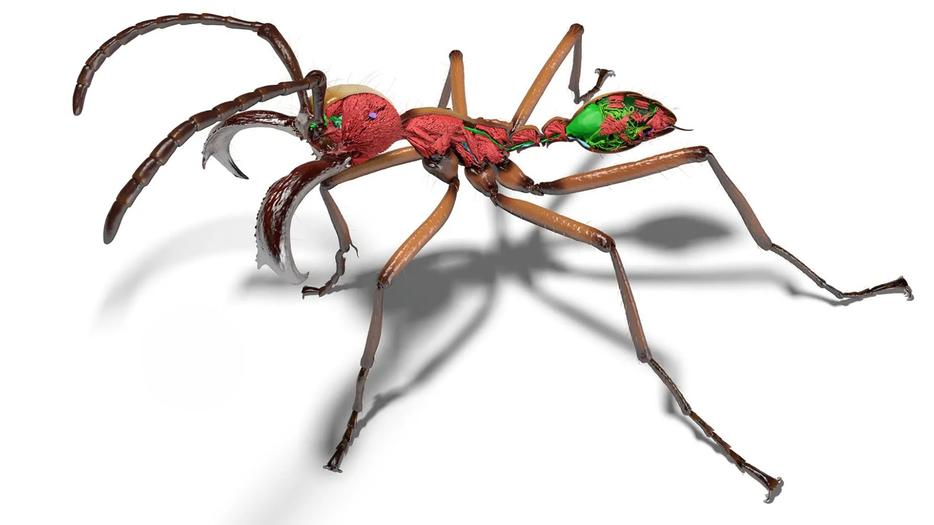
The resulting Antscan models reveal internal details such as muscles, nervous systems, digestive organs, and stingers with micrometer level resolution. These digital ants can also be animated or placed into virtual reality environments for scientific research, education, or entertainment.
“To do this manually would have taken years, so without these computational tools it basically would never have been done,” Economo said. “Now, we are making large strides toward creating a living library of interactive models corresponding to Earth’s biodiversity. AI will enable us to explore the diversity of life and share it with the world.”
Antscan Data Fuels New Research
The growing Antscan database has already proven useful for scientific studies. Economo also served as senior author on a paper published in the journal Science Advances on December 19, 2025. In that research, scientists used Antscan data to investigate whether ant colonies benefit more from having many smaller workers or fewer individuals with stronger bodies.
The team examined relationships between cuticle volume, colony size, and evolutionary diversification across more than 500 ant species. The cuticle forms the protective outer layer of an ant’s exoskeleton. Because producing it requires nitrogen and other minerals, thicker armor represents a larger resource investment for each individual ant.
Their analysis revealed a strong negative correlation between cuticle volume and colony size. In other words, colonies that invest less in thick armor may be able to support more workers, potentially allowing them to grow larger and diversify more successfully.
Antscan made these measurements possible because the 3D models allow researchers to precisely calculate cuticle volume, something that was difficult to measure before. The project also scanned the same ant species examined in a June 2025 study published in the journal Cell and co authored by Economo that generated a set of high quality ant genomes. Together, these datasets could help scientists better understand connections between physical traits and genetic variation.
Because the scans are so detailed, they may also be useful for training machine learning systems to recognize ants in the field during behavioral studies. Economo plans to continue expanding the database by scanning additional specimens and collaborating with UMD computer science students to apply these AI techniques to new biological datasets.
“This work moves us further into the big data era of capturing, analyzing and sharing organismal shape and form,” Economo said. “The potential for integrating these data with other data types and technologies is immense and very exciting.”
Their paper, “High-throughput phenomics of global ant biodiversity,” was published in the journal Nature Methods on March 5, 2026.
This article was adapted from text provided by the Okinawa Institute of Science and Technology.
This research was supported by the German Ministry for Research and Education; the Ministry of Science, Research and the Arts Baden-Württemberg; the German Research Foundation (Grant Nos. INST 35/1503-1 FUGG and 502787686); the Okinawa Institute of Science and Technology Graduate University; the Japan Society for the Promotion of Science (Grant Nos. 18K14768, 21K06326 and 22KJ3077); the Australian Research Council (Award No. IC 180100008); HUN-REN Hungarian Research and the National Research, Development, and Innovation Fund (Grant No. K 147781); the Conselho Nacional de Desenvolvimento Científico e Tecnológico (Grant No. 301495/2019-0); the Critical Ecosystem Partnership Fund, a joint initiative of l’Agence Française de Développement, Conservation International, the European Union, the Global Environment Facility, the government of Japan and the World Bank; the U.S. National Science Foundation (Grant Nos. DEB-1932467, DEB 1927161 and IOS-2128304); the Italian Ministry for University and Research; the Environment and Conservation Fund in Hong Kong (Award No. Nb. ECF 137/2020); and Fundação para a Ciência e a Tecnologia. This article does not necessarily reflect the views of these organizations.
Source link
-
-
-

-

-