In an evolving health landscape, emerging research continues to highlight concerns that could impact everyday wellbeing. Here’s the key update you should know about:
New research reveals how hypoxia-driven red blood cell adaptations may reshape glucose regulation, offering fresh insight into diabetes biology and potential therapeutic strategies.
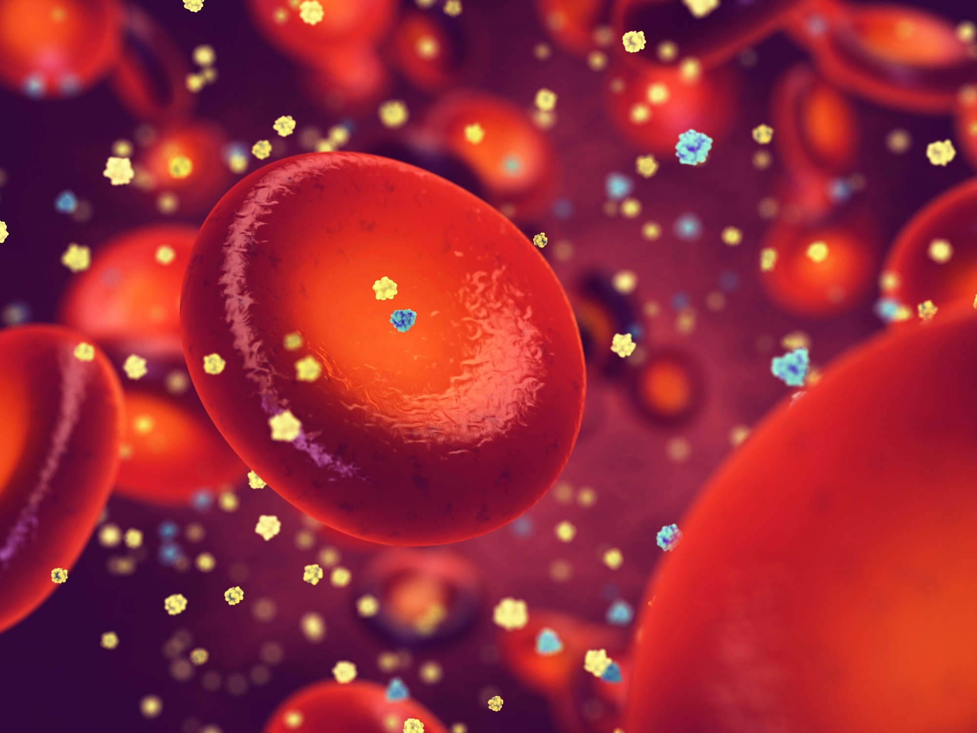
Study: Red blood cells serve as a primary glucose sink to improve glucose tolerance at altitude. Image Credit: nobeastsofierce / Shutterstock
In a recent study published in the journal Cell Metabolism, researchers investigated whether red blood cells (RBCs) function as a primary glucose sink under hypoxic conditions and thereby improve systemic glucose tolerance.
High-Altitude Hypoxia and Improved Glucose Control
Epidemiological observations show that populations living above 3,500 meters exhibit lower rates of diabetes compared to those at sea level. Across Tibet, Peru, the United States, and Nepal, high-altitude communities consistently demonstrate lower fasting glucose levels and improved glucose tolerance. Even animals adapted to altitude display similar metabolic patterns. Despite reduced oxygen availability at high elevations, blood glucose regulation appears enhanced, creating a physiological paradox.
Short-term hypoxia is known to stimulate glucose uptake in peripheral tissues; however, these effects are transient. The persistence of improved glucose control during chronic hypoxia suggests a deeper systemic adaptation. The biological mechanism underlying this sustained effect remained unclear, prompting investigation into whether RBCs contribute directly to whole-body glucose disposal.
Normobaric Hypoxia Mouse Model Design
To isolate the impact of oxygen deprivation, researchers used normobaric hypoxia models in eight-week-old male mice. Animals were maintained in either normoxic conditions (21% oxygen) or hypoxic environments (8% oxygen, equivalent to altitudes above 5,000 meters) for up to three weeks. Blood glucose, body weight, glucose tolerance tests, and insulin tolerance tests were monitored longitudinally.
To determine whether increased RBC abundance influenced glycemia, investigators used two complementary strategies. Serial phlebotomy removed 15% of the total blood volume every three days to reverse hypoxia-induced erythrocytosis. In parallel experiments, packed RBCs from hypoxic or normoxic donor mice were transfused into normoxic recipients.
Glucose uptake was assessed using 2-deoxy-2-[18F] fluoro-D-glucose positron emission tomography/computed tomography imaging and stable isotope tracing with uniformly labeled carbon-13 glucose and carbon-13 2-deoxy-D-glucose. Liquid chromatography-mass spectrometry quantified plasma glucose and intracellular metabolites. Flow cytometry evaluated glucose transporter 1 (GLUT1) and glucose transporter 4 (GLUT4) abundance in RBCs. Proteomic and imaging approaches examined glycolytic enzyme localization and interactions with band 3 protein under varying oxygen conditions.
Hypoxia Rapidly Lowers Blood Glucose Independently of Insulin
Chronic hypoxia significantly reduced basal blood glucose levels within two days of exposure. Glucose tolerance improved at 1, 2, and 3 weeks and persisted for more than a month after mice returned to normoxia. In contrast, insulin sensitivity did not improve and was transiently reduced during hypoxia. The authors interpreted this reduction as a compensatory response to sustained hypoglycemia rather than enhanced insulin action.
Moderate hypoxia (11% oxygen) and intermittent hypoxia similarly improved fasting glucose and glucose tolerance, suggesting potential translational relevance. Hepatic gluconeogenesis did not account for reduced blood glucose levels, indicating increased glucose disposal rather than decreased production was responsible for the observed hypoglycemia.
Red Blood Cells Identified as the Primary Glucose Sink
Whole-body imaging revealed that classical glucose-consuming organs, such as muscle, liver, heart, and brain, accounted for only a minority of increased glucose uptake under hypoxia. This finding suggested the presence of another major glucose-consuming compartment.
During chronic hypoxia, RBC numbers nearly doubled. When erythrocytosis was reversed by serial phlebotomy, blood glucose levels normalized, but improvements in glucose tolerance disappeared. Conversely, transfusion of RBCs from hypoxic donors into normoxic mice induced hypoglycemia without hypoxia exposure. These experiments demonstrated that increased RBC abundance was both necessary and sufficient to drive hypoxia-associated hypoglycemia in this model.
Enhanced Per-Cell Glucose Uptake and Transporter Expression
Beyond increased cell number, individual RBCs under hypoxia exhibited enhanced glucose uptake capacity. Stable isotope tracing showed faster intracellular accumulation of phosphorylated 2-deoxy-D-glucose. Ex vivo experiments confirmed approximately a 2.5-fold increase in glucose uptake per cell.
Flow cytometry revealed upregulated expression of GLUT1 and GLUT4 in hypoxic RBCs. Biotin-labeling experiments indicated that newly synthesized RBCs contributed substantially to the increased GLUT1 abundance, suggesting that erythropoiesis under hypoxia generates metabolically adapted RBC populations.
Metabolic Rewiring Through the Luebering-Rapoport Shunt
Metabolomic tracing demonstrated glucose flux in hypoxic RBCs was redirected toward 2,3-diphosphoglycerate production via the Luebering-Rapoport shunt. Both levels and isotopic labeling rates of 2,3-diphosphoglycerate were elevated. This adaptation enhances oxygen release from hemoglobin to tissues while increasing glucose consumption. The authors noted that precise quantitative flux measurements would require additional targeted analyses.
Low oxygen conditions displaced glyceraldehyde-3-phosphate dehydrogenase (GAPDH) from its inhibitory binding to the band 3 membrane protein, thereby increasing glycolytic flux. This molecular mechanism provided a structural explanation for accelerated glucose metabolism in RBCs under hypoxia.
Therapeutic Implications in Diabetes Models
Hypoxia exposure and hypoxic RBC transfusion improved hyperglycemia in mouse models of type 1 diabetes, enhancing glucose tolerance despite insulin deficiency. In a high-fat diet model of type 2 diabetes, treatment with a pharmacologic agent (HypoxyStat) that increases hemoglobin oxygen affinity and induces tissue hypoxia improved glycemia and glucose tolerance without direct RBC transfusion.
These findings suggest targeting RBC metabolism or safely mimicking hypoxia-induced erythrocyte adaptations may offer therapeutic approaches for hyperglycemic conditions.
Red Blood Cells as Regulators of Systemic Glucose Metabolism
This study identifies RBCs as previously unrecognized regulators of systemic glucose metabolism. Hypoxia increases RBC production and enhances per-cell glucose utilization, enabling RBCs to act as a significant glucose sink independent of insulin signaling. By metabolizing glucose through glycolysis and the Luebering-Rapoport shunt, RBCs improve oxygen delivery and reduce circulating glucose levels.
The findings expand understanding of whole-body glucose homeostasis and suggest potential therapeutic strategies for type 1 and type 2 diabetes. Modulating RBC metabolism or harnessing hypoxic adaptations could represent innovative avenues in metabolic disease management.
-

-

-

-

-