In an evolving health landscape, emerging research continues to highlight concerns that could impact everyday wellbeing. Here’s the key update you should know about:
A large first-trimester study found little evidence that SARS-CoV-2 commonly infects placental tissue, but it also showed that even limited viral presence can reshape immune signaling at the site where pregnancy begins.
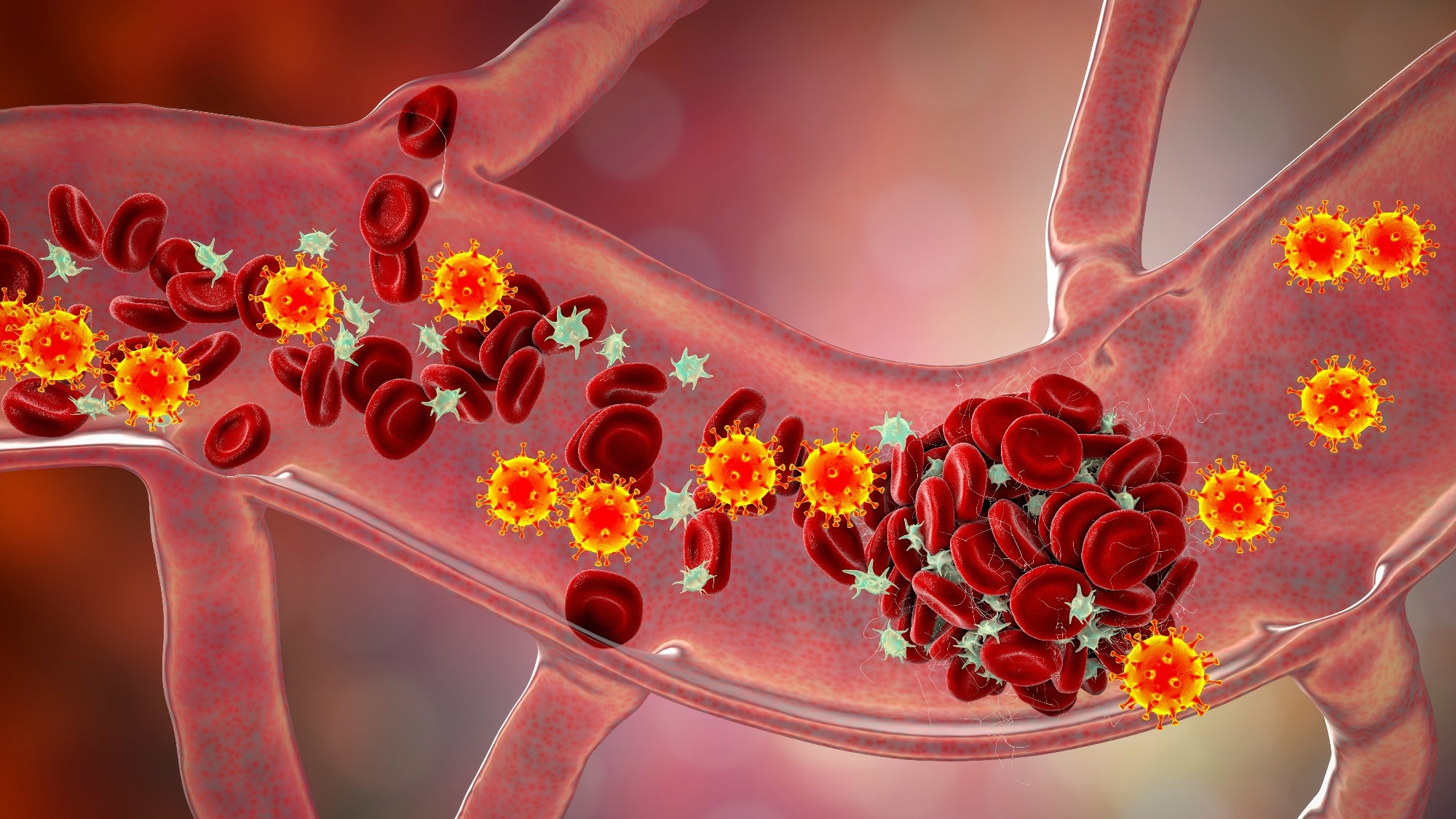
Study: SARS-CoV-2 infection during the first trimester leads to profound immune dysregulation at the maternal-fetal interface despite limited virus detection in placental tissues. Image Credit: Kateryna Kon / Shutterstock
A recent study published in the journal Nature Communications suggests that severe acute respiratory syndrome coronavirus 2 (SARS-CoV-2) is rarely detected in first-trimester placental tissues, suggesting that efficient in utero transmission is unlikely during early pregnancy, despite triggering notable cellular and immune changes in the placenta.
An analysis of 761 first-trimester samples suggested that in utero transmission during early pregnancy is uncommon but revealed placental alterations likely driven by antiviral responses that may affect trophoblast function.
The study also found that higher serum IgG antibody levels were inversely correlated with tumor necrosis factor-beta (TNF-β), suggesting that adaptive immunity may help regulate inflammatory responses and supporting the authors’ view of the potential value of preconception vaccination.
COVID-19 Pregnancy and Placental Risk Background
Coronavirus disease 2019 (COVID-19), now transitioning toward endemic circulation, remains a global health concern. Pregnant individuals are particularly susceptible to respiratory infections due to physiological changes.
Studies have linked COVID-19 to adverse outcomes, including hospitalization, preterm birth, and stillbirth. Although viral components have been detected in placental tissue, confirmed intrauterine transmission appears rare, and its timing remains unclear. Further research is needed to clarify the effects of infection during early pregnancy and its implications for maternal–fetal immune dynamics.
First-Trimester Placental Study Design
About the study. In the present study, researchers investigated the impact of COVID-19 on the maternal-fetal interface in early pregnancy in individuals undergoing elective surgical pregnancy termination within 13 weeks of gestation. They obtained paired decidual and villous tissue samples from the participants between January and September 2023.
The study period spanned two COVID-19 infection waves following the end of China’s strict COVID-19 containment policy, when earlier undetected infections were considered unlikely. To minimize confounding from prior immunity, the investigators excluded participants vaccinated within the preceding 6 months or those with prior severe SARS-CoV-2 infection, based on antibody half-life data.
The team performed reverse transcription-quantitative polymerase chain reaction (RT-qPCR) targeting SARS-CoV-2 envelope (E), nucleocapsid (N), and ribonucleic acid-dependent ribonucleic acid polymerase (RdRp) genes. The analysis also measured host entry genes, angiotensin-converting enzyme 2 (ACE2) and transmembrane protease, serine 2 (TMPRSS2).
Fluorescence in situ hybridization (FISH), along with immunofluorescence assays (IFA), supported the presence of very low levels of viral material rather than a clear active placental infection.
Histological examinations compared decidual and villous tissues from SARS-CoV-2-positive and -negative samples, along with evaluation of host gene expression.
The researchers performed serological analyses to measure anti-receptor-binding domain (RBD) immunoglobulin G (IgG) and IgM levels using chemiluminescence assays. They also conducted cytokine profiling and serum metabolomics to assess systemic immune responses.
To further characterize molecular alterations, the team applied ribonucleic acid sequencing (RNA-seq) at both the tissue and single-cell levels to selected decidual and villous pairs. Subsequently, they conducted differential gene expression (DEG), Gene Ontology (GO), Kyoto Encyclopedia of Genes and Genomes (KEGG), and gene set enrichment analyses (GSEA).
In addition, the investigators analyzed serum samples using ultra-high-performance liquid chromatography-tandem mass spectrometry (UHPLC-MS)-based metabolomics. Lastly, they conducted Pearson correlation analyses to explore associations between antibody levels, inflammatory markers, cytokines, chemokines, and metabolites.
Placental Immune Dysregulation and Low Viral Detection
Results Molecular analyses showed minimal SARS-CoV-2 presence in early placental tissues, with only low-level N gene signals detected in three samples and no evidence of E or RdRp genes, suggesting fragmented or incomplete viral genomes.
RNA-seq also failed to detect viral transcripts, supporting a very low viral burden. Histological assessment showed no structural differences between virus-positive and negative tissues. Although genes for viral entry into host cells (TMPRSS2 and ACE2) were expressed, their limited co-expression across cell types may explain the rarity of placental infection.
Serological analysis of 433 participants classified individuals into acute (IgM-positive), convalescent (IgG-positive), and uninfected groups. Acute infection was associated with elevated pro-inflammatory cytokines, including interleukin-31 (IL-31) and growth-regulated oncogene-alpha (GRO-α).
Contrastingly, the convalescent phase samples showed increased anti-inflammatory IL-10 levels. Notably, IgG levels inversely correlated with tumor necrosis factor beta (TNF-β), suggesting a protective role in immune regulation, whereas IgM levels correlated with multiple inflammatory markers, reflecting active immune responses.
Despite a relatively subdued systemic inflammatory profile, transcriptomic analyses indicated marked local immune activation at the maternal–fetal interface. RNA-seq analyses, together with immune deconvolution and single-cell profiling, indicated increased immune cell infiltration, upregulation of interferon-stimulated genes (ISGs), and increased M2-like macrophage populations.
In addition, disrupted intercellular signaling, particularly involving Wingless-related integration site (WNT) and transforming growth factor-beta (TGF-β) pathways, and downregulation of angiogenesis-related genes were observed. These alterations may impair trophoblast function and placental development, which the authors suggest could potentially contribute to adverse pregnancy outcomes despite limited viral presence.
Preconception Vaccination and Pregnancy Implications
The study findings suggest that SARS-CoV-2 was rarely detected in first-trimester placental tissue, supporting the view that in utero transmission during early pregnancy is uncommon. However, maternal infection was associated with changes in placental immune activity and trophoblast function, which could potentially influence pregnancy outcomes.
The inverse relationship between IgG levels and TNF-β indicates a protective role of adaptive immunity, and the authors say this supports the importance of vaccination before conception. As most cases were mild or asymptomatic, the effects of severe disease may be underestimated.
The authors also note that infection timing was not precisely known and that single-cell RNA sequencing was performed on only one tissue pair per group. Further research should examine long-term outcomes, infection timing, and larger, diverse cohorts to better define maternal and fetal risks.
Journal reference:
- Liu, X. X., Shen, X., Cui, X. et al. (2026). SARS-CoV-2 infection during the first trimester leads to profound immune dysregulation at the maternal-fetal interface despite limited virus detection in placental tissues. Nature Communications. DOI: 10.1038/s41467-026-71770-9, https://www.nature.com/articles/s41467-026-71770-9