

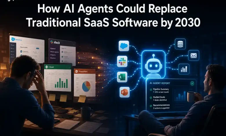
The Death of SaaS: How AI Agents Could Replace Traditional Software by 2030
Imagine a future where you never have to “log in” to work.
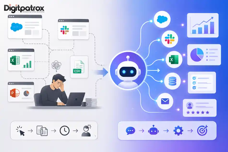
Today, a typical knowledge worker spends their day in a state of digital exhaustion. According to research published by the Harvard Business Review, employees switch between different applications and windows more than 1,100 times a day. We open Salesforce to update a lead, jump to Slack to notify a team, export a CSV to Excel, and manually sync those numbers into a PowerPoint deck for a status meeting.
We aren’t doing deep work; we are performing click-labor.
That shift-from humans operating software to software operating itself-is what is increasingly becoming the Post-SaaS Era. For twenty years, Software-as-a-Service has thrived by selling seats and interfaces. However, the rise of AI Agents-autonomous systems that move from chat to execution-is turning the traditional software dashboard into a bottleneck. We are moving from a world of humans using software to a world of software using software.
⚡ The Executive Summary: At a Glance
The Core Thesis: The value of software is shifting from the Interface (how it looks) to the Agentic Outcome (what it achieves). If your software requires a human to click a button to get a result, you are already legacy.
-
The Problem: SaaS sprawl has created “Integration Debt,” leading to the Death of the Browser Tab as we know it.
-
The Shift: From GUI (Graphical User Interface) to LUI (Language User Interface) and finally HUI (Headless User Interface).
-
The Winner: Platforms that treat APIs as their primary product, not their secondary feature.
📊 Key Highlights for the Modern C-Suite
1. The Death of the “Per-Seat” Model
For decades, SaaS revenue was tied to human headcount. In the Post-SaaS era, this model collapses. As AI agents replace human “clickers,” companies will refuse to pay for seats that no one sits in.
-
The Pivot: Software vendors must move to Outcome-Based Pricing. This marks the end of cognitive debt as businesses stop paying for the process and start paying for the result.
2. Integration Debt vs. Agentic Flow
Most enterprises are buried under “Integration Debt”-the cost of trying to make 100+ apps talk to each other. AI agents act as the universal translator, navigating between tools autonomously.
-
The Pivot: The most valuable asset is no longer the “feature set,” but the accessibility of data via Vector SEO and retrieval windows.
3. The Rise of “Headless” Enterprise
We are entering the era of the Headless Enterprise, where the most important interactions happen in the background via machine-to-machine calls.
-
The Pivot: CTOs should prioritize machine-readability. This transition is being powered by new AI Operating Systems that favor background orchestration over human-clickable buttons.
I. Why Companies Want Fewer SaaS Tools
The shift from traditional software to agentic workflows is happening because companies are hitting a “SaaS Ceiling.” Organizations now manage an average of 130+ SaaS applications.
-
The End of Coordination Work: Knowledge workers spend nearly 60% of their time on “work about work”-communicating about tasks rather than doing them.
-
The Future of Discovery: As software becomes invisible, even the way we find tools is changing through the future of AI search.
II. Companies Already Moving Toward Agentic Workflows
The transition is already observable in the product roadmaps of the world’s largest tech entities:
-
Salesforce Agentforce: Automating CRM workflows without human reps.
-
Microsoft Copilot Studio: Building internal agents across the entire Microsoft 365 ecosystem.
-
Claude “Computer Use”: Anthropic’s latest capability allows AI to see a screen and move a cursor. This is part of the new Claude AI handoff workflow designed for seamless automation.
III. Traditional SaaS vs. AI Agentic Workflows
To understand the business impact, we must distinguish between simple triggers and true intelligence.
| The Software Operator (Today) | The AI Supervisor (Tomorrow) |
| Tool: Traditional Automation (Zapier). | Tool: AI Agents. |
| Logic: Linear “If-This-Then-That.” | Logic: Reasoning, Planning, and Execution. |
| Failure Mode: Breaking on UI changes. | Failure Mode: Hidden Decay/Prompt Rot. |
| Status: Digitally Exhausted. | Status: Strategically Focused. |
IV. Why Most AI Agent Startups Will Fail
Despite the hype, replacing SaaS is harder than it looks. Businesses don’t just buy software functionality; they buy accountability.
The winners of the Post-SaaS era will be those that solve the trust gap through AI Reliability Engineering. For a CEO, an agent that occasionally makes invisible mistakes is far more dangerous than a slow human. Furthermore, as agents become the primary users of software, AI Cybersecurity will become the most critical component of the enterprise stack.
V. The Hidden Risks: “Prompt Rot”
As organizations move from standardized SaaS to custom Agentic Workflows, they face Prompt Rot. Unlike a SaaS platform updated by Google, a custom agent’s logic is often frozen in a specific prompt. When the underlying model is updated, the agent’s behavior may drift, leading to silent errors.
VI. CTO/CEO Action Plan: Preparing for 2030
-
Prioritize AX (Agent Experience): Stop buying software based on how the UI looks. Start buying based on the quality of the API.
-
Audit for “Click-Labor”: Identify which departments spend more than 20% of their time on data entry. Small businesses are already saving 10+ hours weekly by automating these specific frictions.
-
Kill the “Seat” Model: Negotiate contracts based on outcomes rather than licenses.
🚀 The Bottom Line
For decades, humans adapted themselves to software interfaces. The Post-SaaS era flips that relationship: software adapts itself to humans.
The next decade of computing may not be defined by better dashboards or more apps. It may be defined by the disappearance of the interface itself. The winners won’t necessarily be the companies with the most features, but the ones that remove the most friction between intention and execution.
VII. SERP Capture Layer
-
5 FAQ Questions:
-
Is SaaS dying? (A: The seat-based, UI-heavy model is declining in favor of outcome-based AI agents).
-
Will AI replace Salesforce? (A: Salesforce is pivoting to an agent-first model with Agentforce to remain the core infrastructure).
-
What is the Post-SaaS Era? (A: A shift where AI agents operate software on behalf of humans).
-
What is Prompt Rot? (A: The performance decay of AI agents as underlying models change over time).
-
What is click-labor? (A: The repetitive, manual task of moving data between different software applications).
-
-
Meta Description: Explore why AI agents are replacing traditional SaaS dashboards. Learn about the Post-SaaS era, the UI-Utility Inversion, and how companies are shifting from operating software to directing outcomes.
-
Internal Anchor Text: “Learn more about the difference between AI agents vs. traditional automation and how to manage system decay.”